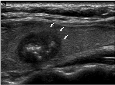
abnormal thyroid cancer ultrasound colors
Seroma hematoma abscess tumors. A pictorial review hot wwwncbinlmnihgov.

Pin On Abd 300 Mod 4 Ultrasound
Ad Memorial Sloan Kettering Has Experts for Every Type of Cancer Including Yours.

. The thyroid gland was evaluated for any nodules following carotid Doppler ultrasound in 290 patients. A common imaging test used to evaluate the structure of the thyroid gland. The probe detects these reflections to make pictures.
Ad Learn what causes thyroid nodules and the complications that can arise. We identified it from well-behaved source. A solid one is more likely to have cancerous cells but youll still need more tests to find out.
The most prevalent form of thyroid cancer is papillary thyroid cancer 75-80 followed by follicular 10-20 medullary 3-5 and anaplastic 1-2 thyroid cancers 2 26. Ad Learn more about the signs that may reveal you have an Issue that need attention. Normal vs abnormal thyroid ultrasound thyroid ultrasound colors meanings.
An ultrasound may show your doctor if a lump is filled with fluid or if its solid. This nodule shown in red comprises about 80 of the thyroid tissue shown in yellow in this particular area of the thyroid. Thyroid hypoechogenicity at ultrasound is a characteristic of autoimmune thyroid diseases with an overlap of this echographic pattern in patients affected by Graves disease or Hashimotos thyroiditis.
World Class Cancer Care Treatment. If you looked at other parts of the thyroid however you would not see the nodulem. It measures 057 x 061 x 069cm.
Patients and methods. Color Doppler showed increased vascularity while the rest of the thyroid showed normal vascularity. We tolerate this kind of Abnormal Parathyroid Ultrasound graphic could possibly be the most trending subject in imitation of we ration it in google plus or facebook.
Thyroid ultrasound reveals slight increase in size of lobes from 3 5x12x9 mm to 41x21x 12mm and isthmus is 1. Thyroid nodule Thyroid cancer Ultrasound Fine-needle aspiration Sonographic featuresIntroduction. Can you detect thyroid cancer in ultrasound.
Color and Power Doppler ultrasound failed to show significant vascularity within the affected area lesion in the right lobe. Buy 2021 Quality Abnormal Thyroid Cancer Ultrasound Color Doppler directly with low price and high quality. Here are a number of highest rated Abnormal Parathyroid Ultrasound pictures upon internet.
She was then referred to me for ultrasound. Color on your thyroid ultrasound means that color doppler was applied and blood flow was detected. After thyroidectomy the local inflammatory response results in proliferation of fibrofatty connective tissue which fills the dead space made by surgery There is also displacement of the strap muscles the carotid sheath structures and the cervical esophagus.
Aim of the present paper was to study the thyroid blood flow TBF by color-flow doppler CFD an. The first links in each row here correspond to ultrasound color post-processed images. Tsh slightly high inspite of synthroidhave hashimotos is the increase abnormal.
Could it be cancer. Ultrasound showed a well-defined rounded isoechoic solid nodule in the upper pole of right thyroid lobe. The normal thyroid gland is located in the anterior lower neck between the thyroid cartilage and the thoracic inlet.
Enlarged mildly hypoechoic heterogeneous. Ultrasound uses soundwaves to create a picture of the structure of the thyroid. Thyroid disorder Grayscale ultrasound Color doppler Key features.
The ultrasound will also show the size and number of nodules on your thyroid. Ultrasonography of thyroid nodules. Enlarged heterogeneous with lobular margins.
The survival rate for thyroid cancer in general is better than for other forms of cancer. Transverse gray-scale ultrasound neck a shows a large well circumscribed oval shaped widthlength hyperechoic nodule in a thyroid lobe. Markedly Markedly hyperemic.
Ultrasound imaging of the thyroid gland shows markedly hypoechoic lesions in the right lobe. Benign thyroid adenoma in a 42-year-old female patient. The hypoechoic thyroid lesion shows irregular borders and is seen to infiltrate along the long axis of the affected lobe.
Hypoechoic and micronodular septal lines. These images are examples of pathology I detect with sonograms. Its submitted by government in the best field.
The lesion has slight heterogeneous appearance due to presence of few tiny cystic spacesclefts. Doctors in 147 specialties are here to answer your questions or offer you advice prescriptions and more. Book an Appointment with the Eperts at MSK.
This page aggregates the highly-rated recommendations for Thyroid Ultrasound Normal. For papillary thyroid cancer the 20-year survival after surgery is around 99. Ultrasound color and grayscale images pictures Ultrasound Color images.
If there was an abnormal finding in the thyroid ultrasound the patient was referred to an endocrinologist and after clinical and laboratory evaluation fine-needle aspiration FNA biopsy was done if required. The color Doppler appearance of each nodule was graded from 0 for no visible flow through 4 for extensive internal flow. Find out the surprising causes and risk factors of thyroid nodules immediately.
Thyroid is a gland that serves several functions that affect the well being of the body. They are the choices that get trusted and positively-reviewed by users. It is generally normal unless there is too much co.
We obtained color Doppler images of thyroid nodules undergoing sonographically guided fine-needle aspiration. Other links to color or gray scale images will be included here later. A thyroid nodule is a discrete lesion within the normal thyroidSuch nodules are a common occurrence in the general population and a frequent incidental finding on.
An abnormal growth of thyroid cells that forms a lump within the thyroid. Send thanks to the doctor. What is the red and blue on a thyroid ultrasound.
You would only see normal thyroid tissue. While most thyroid nodules are non-cancerous Benign 5 are cancerous. A thyroid function test was done and confirmed T4 is elevated.
To determine whether color Doppler interrogation of a thyroid nodule can aid in the prediction of malignancy. Red and blue denote.

Ghim Tren Things That Are Wrong With Me

Pin By Dr Abuaiad On Lymphatics Diagnostic Medical Sonography Nuclear Medicine Sonography

Ultrasound Image Gallery Thyroid Ultrasound Thyroid Diagnostic Medical Sonography


